Biology’s Most Stunning and Strange Images of the Year.
WORLD OF BIOLOGY.
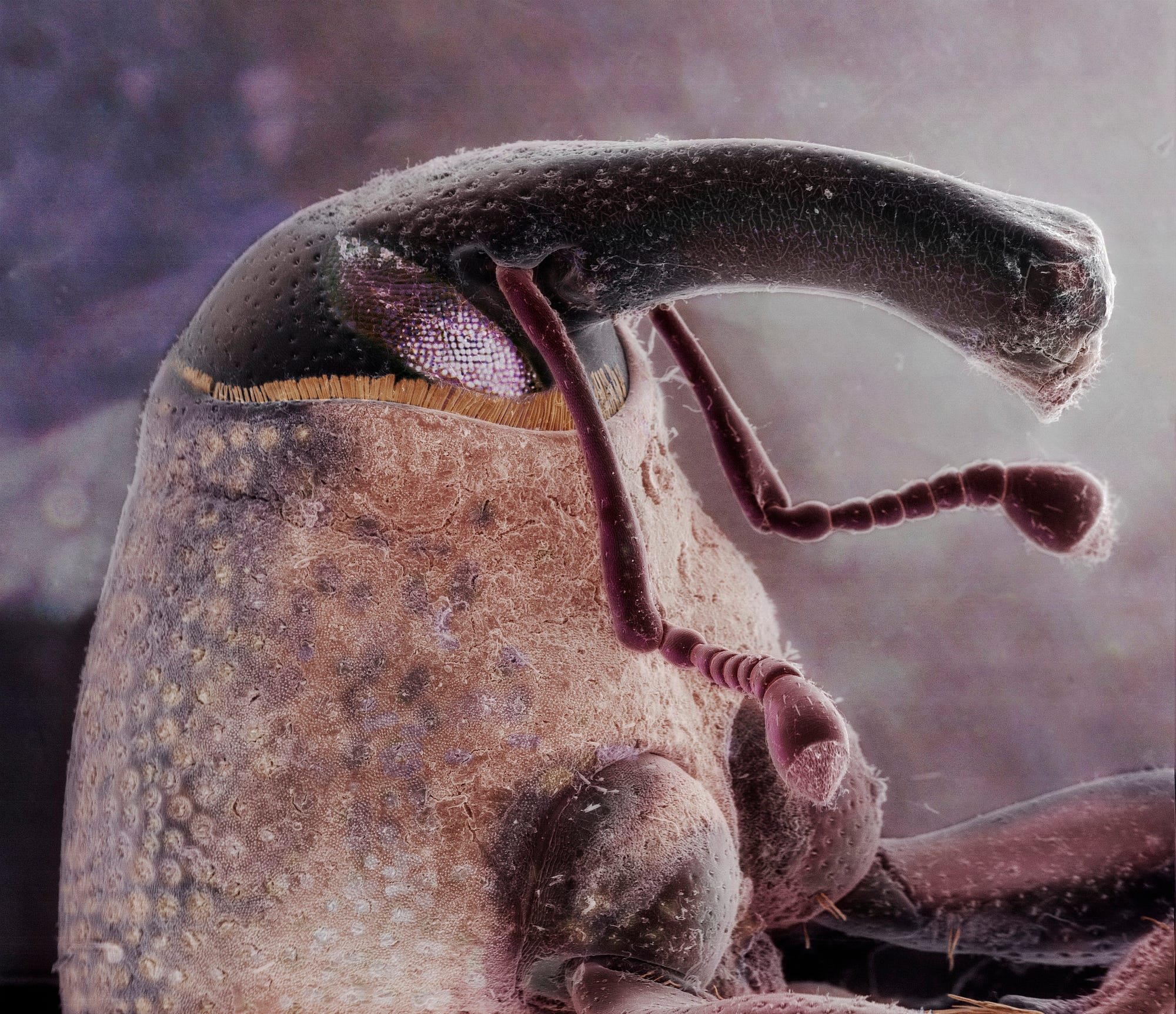
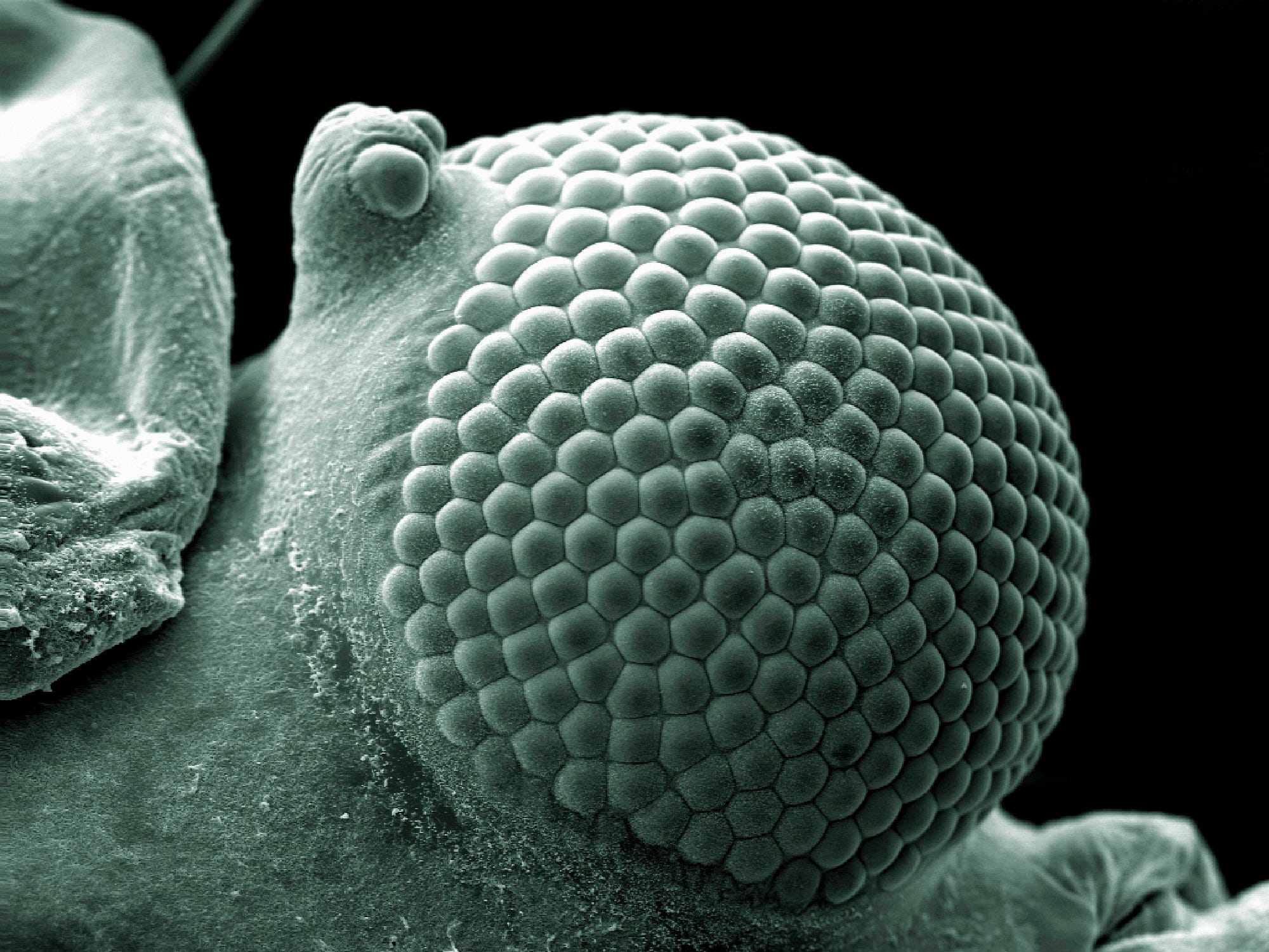
Scanning electron microscope composite image of the head of a boll weevil (Anthonomus grandis) found on the front porch of a suburban house. The boll weevil is a beetle that feeds on and lays its eggs in the cotton plant. These agricultural pests have long curved snouts (often half as long as their bodies) and can destroy entire cotton crops. They develop from egg to adult in approximately 20 days and grow on average to 6–8 mm in length. This is one image in a series of work looking at common household pests found inside homes on the outskirts of town. These images of our often-overlooked housemates are in the style of traditional portraits. The width of the image is 4.1 mm. — Daniel Kariko
Biology’s Most Stunning and Strange Images of the Year
The biomedical image award that shows the surreal beauty of the natural world
Few disciplines are as full of complexity and mystery as biology. But as with any science, public interest has to be earned. Directing public eyeballs toward the study of life is why the UK-based Wellcome Trust hosts the Wellcome Image Awards, presenting some of the most unbelievable, uncanny, and sometimes unnerving images from all of biological science.
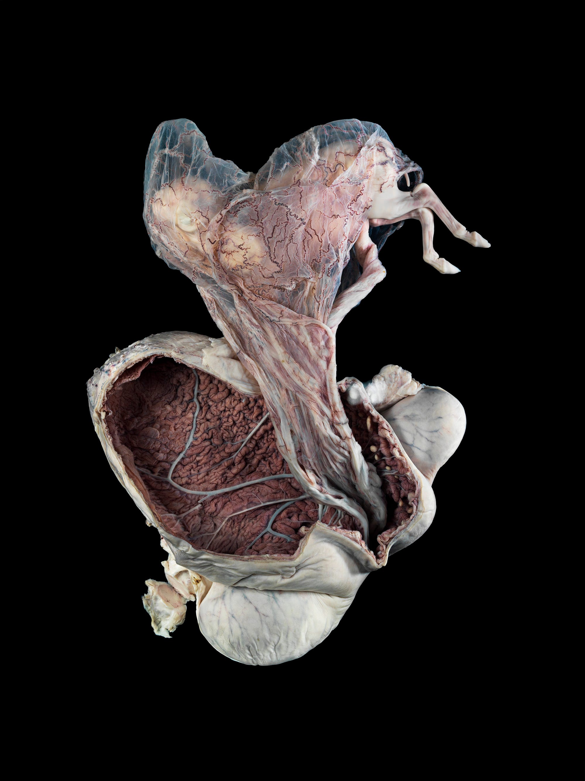
This year’s overall winner, announced today, is a jaw-dropping frame showing a 5-month-old horse fetus. Floating above its opened womb, the delicate organic veil of the placenta parts just enough to reveal two diminutive hooves of a developing foal. It’s a stunning expression of the fragility of life, equally morbid and serene. It won because it meets the explicit aim of the award: to attract the public eye and inspire wonder.
Photograph of a pregnant uterus from a New Forest pony, approximately five months into the pregnancy. The developing pony (fetus) is outside the uterus but remains attached by its membranes and umbilical cord. The bent back legs of the fetus are sticking out from the membranes (top right-hand side). The uterus has been cut open to reveal its vast blood supply, which is visible on the inner surface. This historical specimen is from a cull animal that happened to be pregnant at the time. It is preserved in formalin in a Perspex container and was photographed in the Anatomy Museum of the Royal Veterinary College in London. The container measures 48 x 30 x 7 cm. Pregnant uterus, equine — Michael Frank, Royal Veterinary College.
“It’s not just spreading science literacy, it’s not just like a paternalistic approach where we want to ‘teach the masses about science,’” says Catherine Draycott, Head of Wellcome Images, “It’s more about engaging people and saying, my god this is amazing stuff, what could that possibly be a picture of?,’ and drawing them in.”
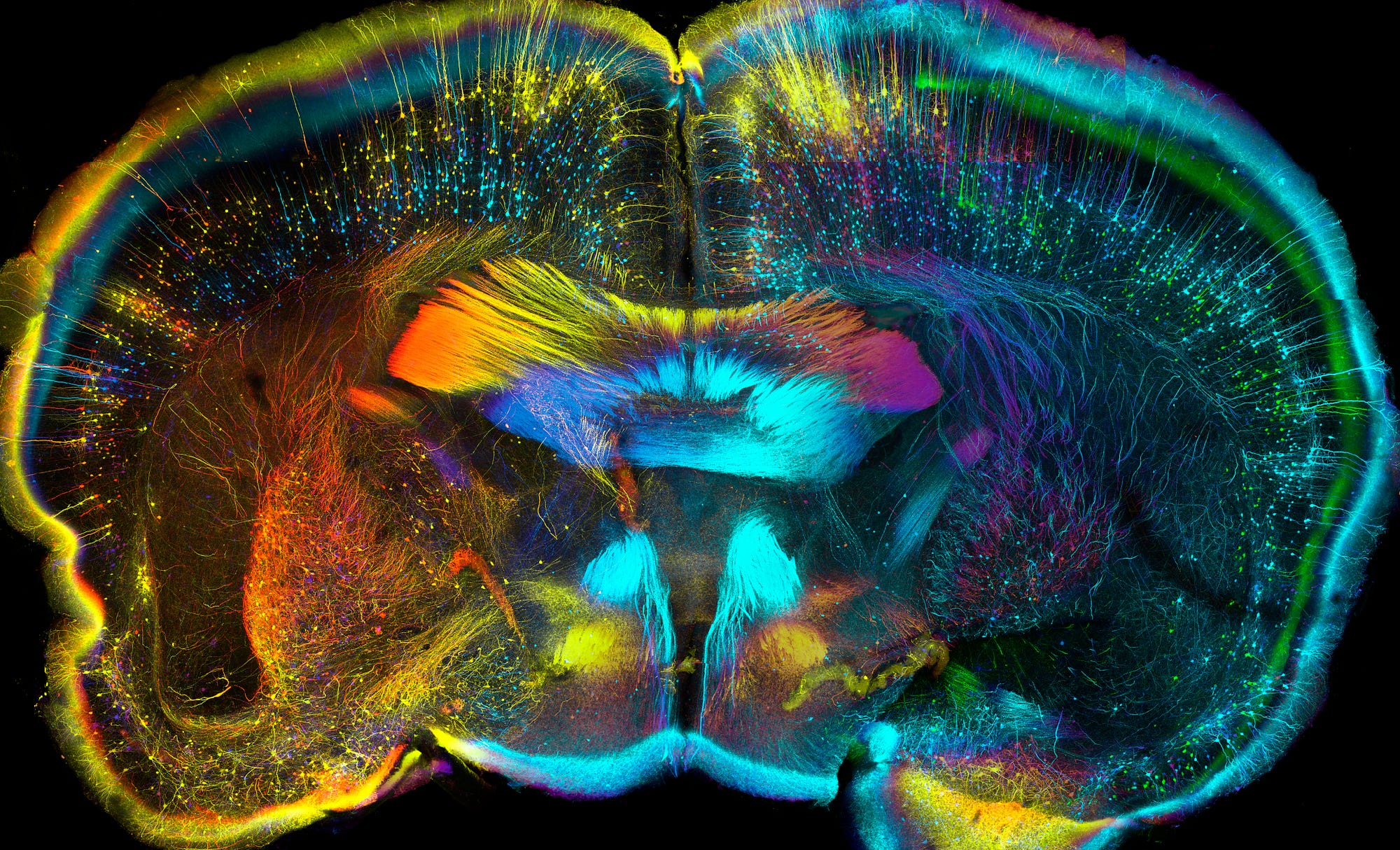
Confocal micrograph of nerve cells inside a section of adult mouse brain. The brain has been sliced (like a loaf of bread), and one of those pieces is seen here. After being sliced, it was chemically treated to make the tissue transparent so that structures deep inside could be more easily seen. A subset of nerve cells tagged with a visual marker (green fluorescent protein) were visualised at different depths through the piece of tissue, which is 0.75 mm thick. The markers are colour coded from red (nearest) to orange, yellow, purple, blue and green as you look into the image. This technique is being used to map the complex wiring of whole brains. This brain is 7.4 mm wide.— Luis de la Torre-Ubieta, UCLA
Other contenders include the cross-section of a cat tongue, a micrograph of a 2mm long Parasitoid wasp, and the back of an elderly woman afflicted with scoliosis. Not all the images are photographs though — included also are an illustration of pollen grains and a 3D-printed scan of a patient’s lungs and rib cage. Anything from hand drawn illustrations to the most advanced imaging technologies are considered. This flexibility in media means that the concepts or facts contained in an image can take center stage over the technical achievement, although the range and complexity of the submissions is definitely on the rise as imaging technology improves.
Illustration of pollen grains being released from a flower in the Asteraceae family. Asteraceae is one of the largest families of flowering plants and is commonly known as the aster, daisy, sunflower or composite family. Pollen grains contain the male sperm cell and are produced in the anther, one of the male parts of the flower. They are carried to other flowers — primarily by insects, birds and the wind — so flowering plants can reproduce. They look like fine dust and are a common cause of hay fever or seasonal allergies. Pollen grains come in all shapes and sizes, but they are usually between 0.01 and 0.1 mm in size. — Maurizio De Angelis
“We do find that the judges tend to be much harder on the straight photography and straight illustrations because they expect so much from fine art photography and illustration,” says Draycott. “With the science images they’re almost willing to accept maybe some blurriness here and there, because it’s the real thing, and they know it is and it’s something you can’t see with the naked eye.”
Cat tongue, cross section — David Linstead
Scanning electron micrograph of a greenfly eye. Many species of aphids, also commonly known as greenfly, are major agricultural pests that feed on plant sap. Aphids have a pair of curved compound eyes that bulge out of the head and have a wide angle of view. Each eye is made up of thousands of repeating units known as ‘ommatidia’, each with a tiny lens on the front surface. Each lens faces a slightly different direction, and together they produce a mosaic image. This allows the fly to see very quick movements but not fine details or objects that are far away. The small circular structure (ocular tubercle; top left) may help insects see polarised light. The width of the image is 280 micrometres (0.28 mm).— Kevin Mackenzie, University of Aberdeen
The award isn’t a competition, and anybody can submit, scientist or not. The winning image of a horse uterus wasn’t taken by a researcher, for example, but by a fine art photographer. In contrast, the sensitively framed and dramatically lit photo of a scoliotic spine was taken as part of a researcher’s regular documentation work.
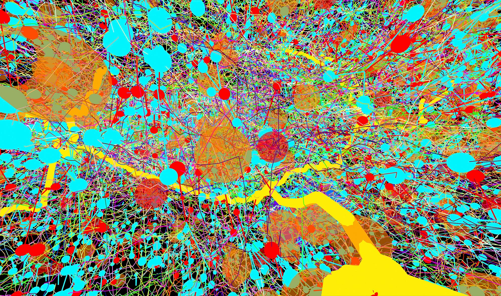
Reminiscent of a Jackson Pollock painting, this image shows part of the central nervous system in a fruit fly (Drosophila melanogaster). Transmission electron micrographs were used to create a digital colour-coded map of the area. An organism’s nervous system controls everything it does, from breathing and moving to thinking and feeling. Instructions to perform these tasks are carried by cells called neurones. A neurone able to sense vibrations (yellow) is surrounded here by lots of other neurones, each depicted as a single line. Messages enter (blue circles) and exit (red circles) neurones at points of contact called synapses. Other features of interest (orange circles), such as mitochondria, are also marked. The width of the image is approximately 15 micrometres (0.015 mm). — Albert Cardona, HHMI Janelia Research Campus
Each image reveals a sublime perspective on things we may usually consider mundane — the fly that’s annoying you as you settle in to read a book is, upon close inspection, a living and rugged alien landscape; a tractographic MRI of the brain is hard to distinguish from a map of global shipping routes, only this one is for thoughts and actions instead of goods and materials. Such synesthetic shifts in perspective are the real power of images like these.
“It’s about understanding life, death, sex, and disease: the cornerstones of drama and art,” said scientist and broadcaster Adam Rutherford, while presenting the awards at this year’s ceremony.
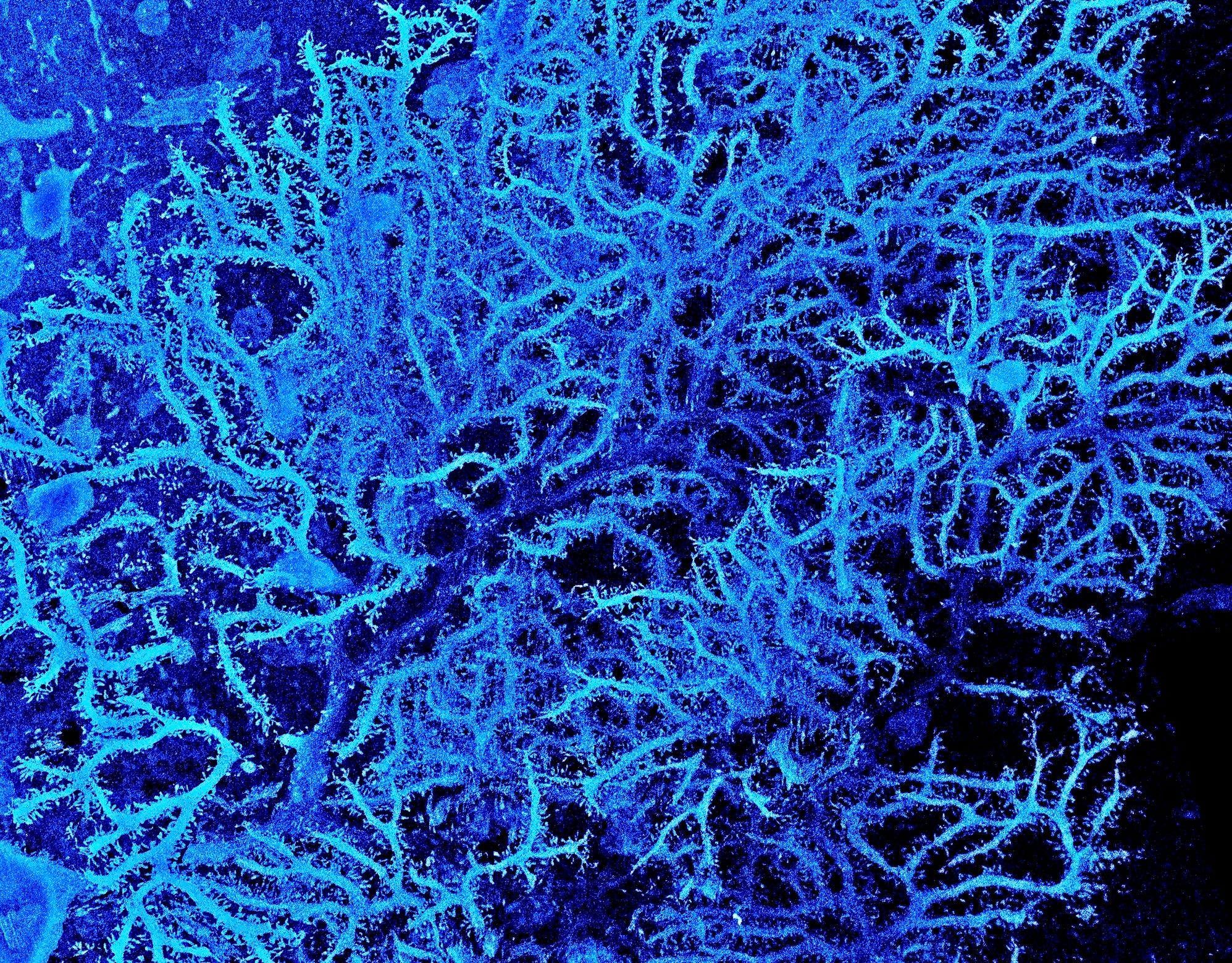
Scanning electron micrograph of tree-like branches (dendritic tree) spreading out from a particular type of nerve cell (Purkinje cell, or neurone) found in the brain. The finger-like projections in this elaborate network act like tiny sensors, picking up information and passing on messages to help control and coordinate muscle movement. This particular neurone is from the cerebellar cortex in a rat brain. To allow us to see the dendritic tree, this Purkinje cell was filled with a visual marker before being imaged by focused ion beam scanning electron microscopy, which allows neurones and neural circuits to be reconstructed in high resolution. The width of the image is 110 micrometres (0.11 mm). — Prof. M. Hausser, Sarah Rieubland, andArnd Roth, UCL
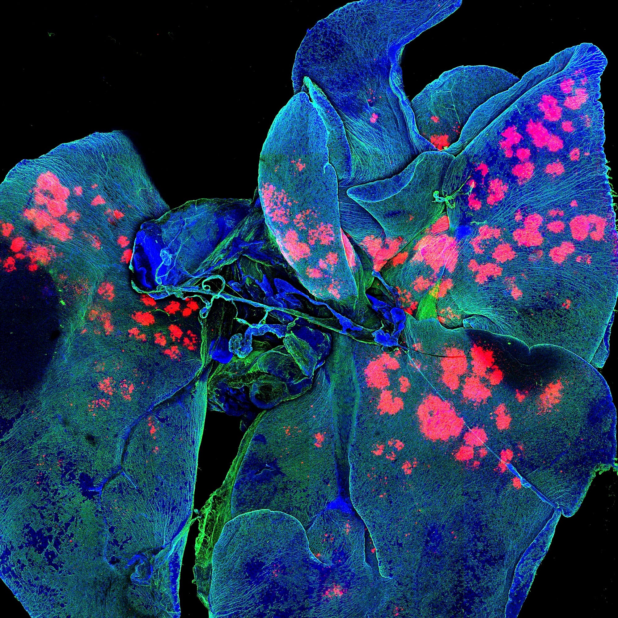
Confocal micrograph of whole mouse lungs (blue and green). Microparticles that can carry medicines (pink) are being studied to see whether they can deliver these drugs to the lungs. Current anticancer therapies have many toxic side-effects, so researchers hope that these microparticles could one day deliver anticancer medicine in a much simpler, more targeted way — for example, in an inhaler — with fewer side-effects. As microparticles release drug over time, fewer doses may be needed. The right lung in mice is divided into four lobes (right side of image), but the left lung has only one lobe (left side of image). Remnants of the windpipe (trachea) and surrounding tissue are also visible (centre). The width of the image is 12.7 mm. — Gregory Szeto, Adelaide Tovar, Jeffrey Wyckoff, Koch Institute, copyright MIT
The Wellcome Trust, established in 1936, has held the award sporadically since 1997 in accordance with the wishes of founder and pharmaceutical magnate Henry Wellcome. He was himself a prodigious collector of medical artifacts, and saw to it that his immense fortune would continue to support medical research. The trust went public in the 80s, and maintains investments all over the world and in countless industries outside of the pharma realm. While Wellcome’s business ended up through various mergers as a part of the mega pharmaceutical company Glasko-Smith-Kline, the Wellcome Trust maintains an independent endowment of nearly $30 billion, making it the second largest private funder behind the Gates Foundation.
As such, it’s well situated to partner with other organizations in promoting medical science. Partnerships with MIT, the University of Texas in Galveston, and numerous other museums and organizations across the UK means images from the award will be on display at 11 such locations, in addition to the exhibition space in Wellcome’s London headquarters. They’ll also join the more than 40,000 images hosted on the Wellcome’s online collection.
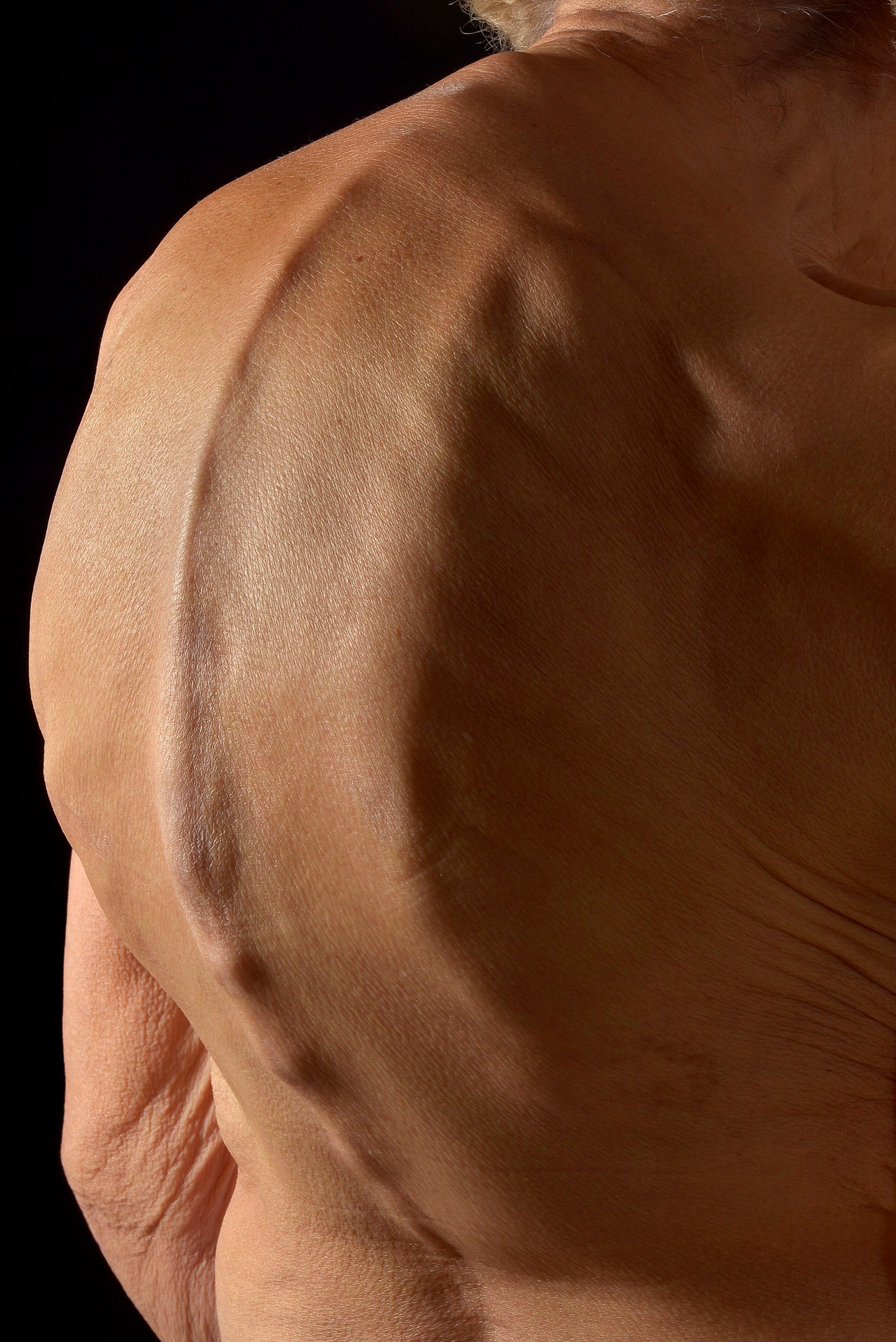
Photograph of a 79-year-old woman’s back, showing an abnormally curved spine. This hunched back appearance is known as kyphosis, or ‘dowager’s hump’, and causes the upper back and shoulders to round forward. Although kyphosis can occur at any age, it is most commonly seen in elderly women. There are many different causes, including poor posture, injury, osteoporosis, cancer and cancer treatments, infection, a birth defect, and degenerative or endocrine diseases. In addition to having an abnormally curved spine, other symptoms can include back pain, stiffness and — in severe cases — difficulty breathing or eating. Treatment options are varied and will depend on the cause and severity of the condition. — Mark Bartley, Cambridge University Hospitals NHS Foundation Trust
Light micrograph of a tiny parasitoid wasp (Wallaceaphytis kikiae) viewed from above. Parasitoid wasps lay their eggs inside other insects. After hatching, the larvae feed on their host, eating it alive from the inside out. This is a new genus of parasitoid wasp recently discovered in the rainforests of Borneo, where a single female wasp was found mixed in with thousands of other insects. It measures only 0.75 mm in length and has unusual antennae, legs and wings. It’s named after Alfred Russel Wallace, who coauthored the first ever publication on evolution by natural selection with Charles Darwin and who himself identified new insects while in Borneo in the mid-19th century. Even today, Borneo is still known to be rich with other undiscovered species. — Andrew Polaszek, Natural History Museum
As the volume of submissions increases and public interest broadens, the Wellcome Image Award may become an annual event. As the work of a charitable organization, it’s encouraging to see a channel opening ever wider for images that inspire curiosity, about a field that needs as much support as it can get. Whether or not they can show us a picture of it, Wellcome’s heart seems to be in the right place.
WORLD OF BIOLOGY.
“It’s not intended to be in any way partisan, it’s intended to be engaging to anyone who might be interested in science or to get people interested in science,” says Draycott. “The idea is that we just want more people interested in order to have more bright young people going into the field.”


0 comments:
Post a Comment
World of Biology.